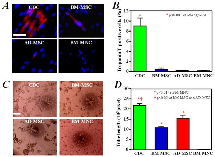
Figure 3. In vitro myogenic differentiation and angiogenesis assay.
A) Troponin T, with distinct myocyte-like appearance, was expressed spontaneously in a fraction of CDCs cultured for 7 days. This cardiac-specific marker was rarely expressed in BM-MSCs, AD-MSCs, and BM-MNCs. B) Quantitative analysis of Troponin T expression (n=3) in CDCs (9% of the cells positive), BM-MSCs (0.4% positive) and AD-MSCs and BM-MNCs (approximately 0.1% positive). C) CDCs, BM-MSCs, and AD-MSCs produced capillary-like tube formations in extracellular matrix. BM-MNCs did not form similar structures under these conditions. D) Quantitation and comparison of tube formation capacity by the different cell types (n=3). Bars = 50 um.

