
Figure 5.
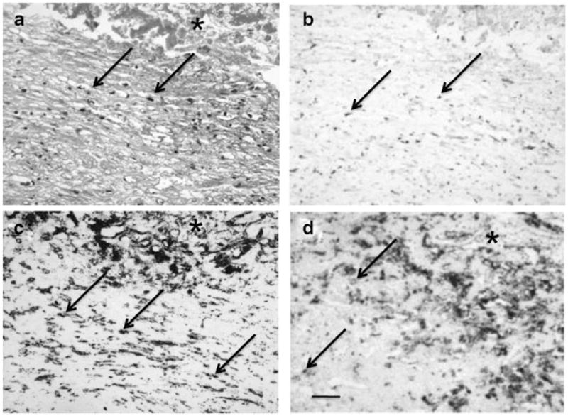
Histopathology of the choroidal neovascular membrane (CNVM) specimens from the second age-related macular degeneration patient who was unresponsive to ranibizumab therapy. (a) The CNVM consists of mild monocytic infiltration (arrows) and hemorrhages (asterisk); (b) Cells in the CNVM express VEGF (arrows); (c and d) Infiltrating cells are CD68+ (c) and CD163+ (d) macrophages. (Original magnifications: ×100, scale bar, 60 μm; a, hematoxylin, b–d, avidin biotin-complex immunoperoxidase).