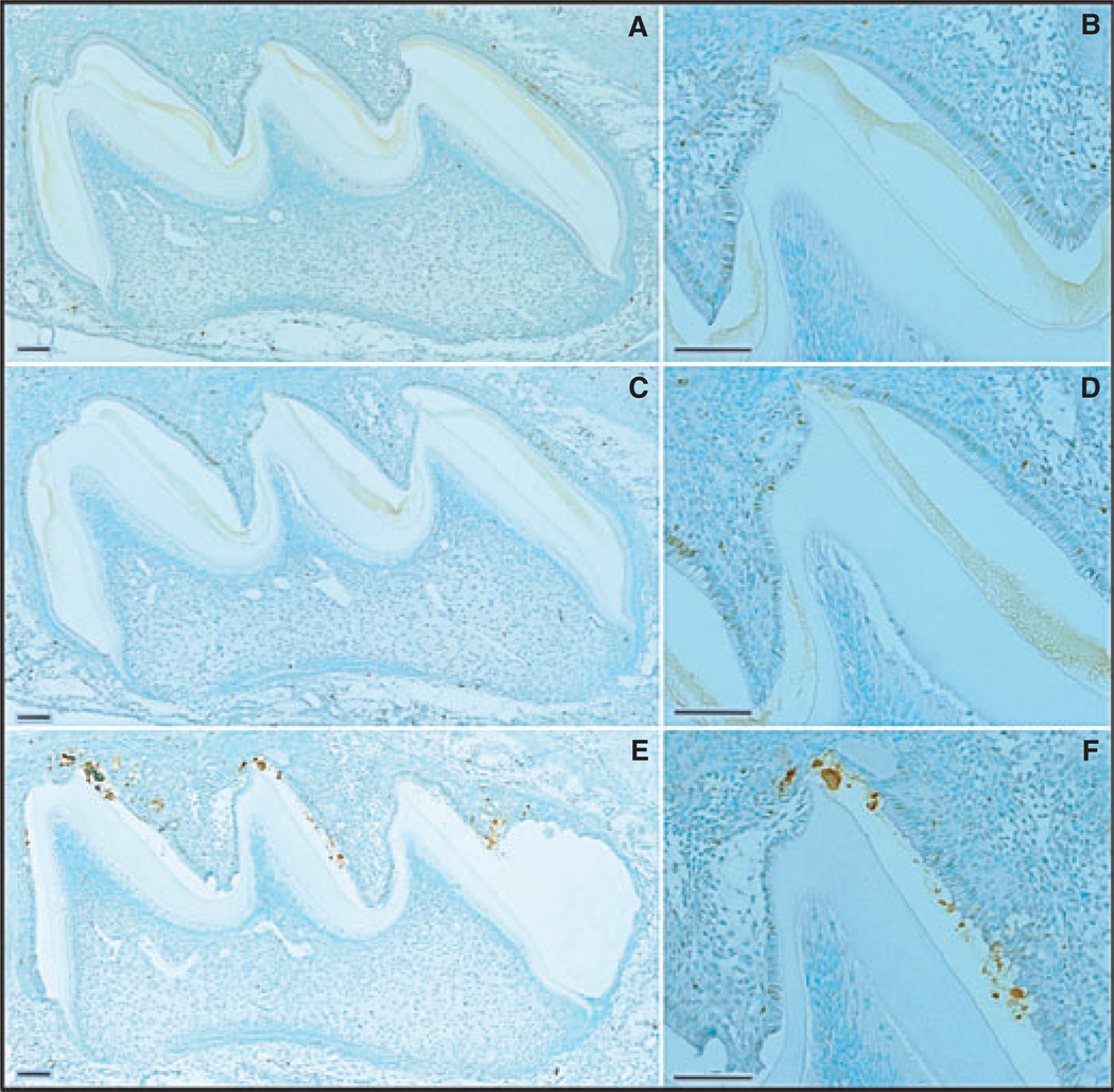
Fig. 3.
Apoptosis [determined using terminal deoxynucleotidyl transferase (TdT)-mediated biotin–dUTP nick-end labelling (TUNEL)] and assessment of day-9 mouse maxillary first molars from wild-type (A, B), enamelin (Enam) heterozygous (C, D), and Enam null (E, F) mice. Panels on the right (B, D, F) detail the apoptotic staining in the ameloblast layer of the central cusp tip. Bars = 100 µm. Ameloblasts are in the maturation stage of amelogenesis.

