Figure 2.

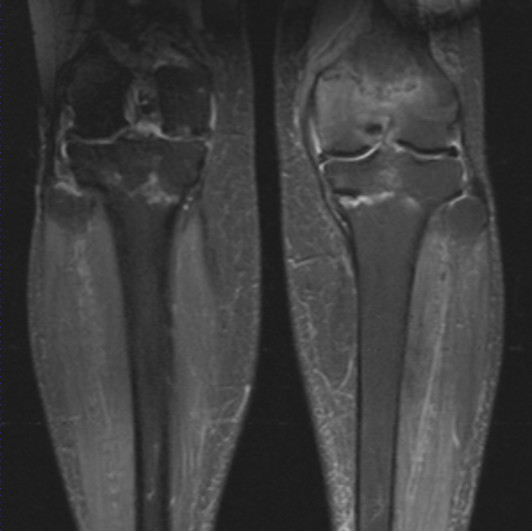
Magnetic resonance imaging coronal short inversion time inversion recovery images show a linear signal change in both the proximal tibia and left distal femur with surrounding reactive change.

Magnetic resonance imaging coronal short inversion time inversion recovery images show a linear signal change in both the proximal tibia and left distal femur with surrounding reactive change.