
Figure 2.
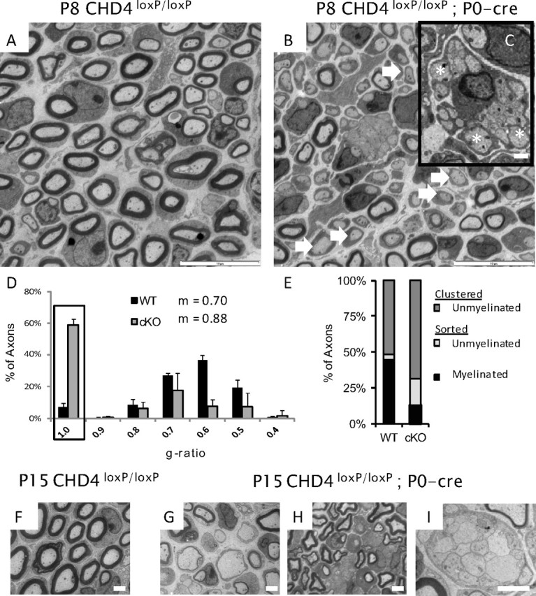
Chd4 mutant mice show a developmental delay. A, B, Electron microscopy analysis of sciatic nerve from wild-type and Chd4 mutant mice at P8, showing hypomyelination (arrows) and a radial sorting defect where large-caliber axons are still associated with Remak bundle axons (C, inset) (asterisks). Scale bars: 10 and 1 μm. D, The g ratio measures the ratio of axon diameter to its myelinated diameter. Unmyelinated Schwann cells are represented with a g ratio of 1 (box). Histogram error is given as mean SD for each bin. E, Axon classification into categories at P8: clustered-unmyelinated (dark gray), sorted-unmyelinated (light gray), or sorted-myelinated (black). P15 WT sciatic nerve (F) contrasts with hypomyelination (G, H) and radial sorting defects (I) in Chd4 mutant nerve. Scale bar, 2 μm.