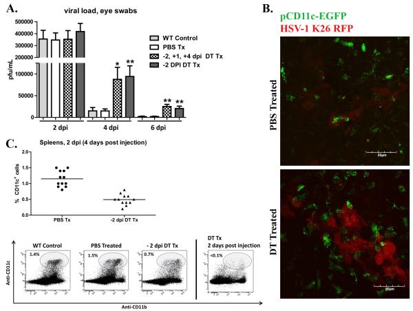
Figure 3. Dendritic cells are required for optimal early clearance of HSV-1 from the cornea.
Wild type (WT) BALB/c mice andCD11c-DTR chimeras were infected with HSV-1-RE. CD11c-DTR chimers were treated with PBS or DT 2 days before infection (−2 dpi); or treated with DT at −2 dpi, +1 dpi, and + 4 dpi. A) At indicated time eyes were swabbed with a sterile surgical spear and assayed for live virus by standard plaque assay. The data are presented as the mean ± SEM viral plaque forming units (pfu) per cornea.B) CD11c-DTR chimeras were treated with PBS or DT 2 days before corneal infection with HSV-1 K26-RFP. At 2 dpi, whole corneas were excised, mounted, and analyzed via confocal microscopy. Representative images depicting virally infected cells (red), and pCD11c-EGFP+ DCs (green) within the viral lesion in the central cornea .C) Spleens were harvested at 4 dpi from WT mice or CD11c-DTR chimeras that received ip PBS or DT treatment 2 days before HSV-1 corneal infection; cells were dispersed, stained with antibodies to CD11c and analyzed via flow cytometry. DCs had partially recovered by 4 days after treatment (2 dpi) but remained significantly reduced relative to PBS treated controls (p<0.01). (** p<0.01, * p<0.05), n=4-12 mice per group, 2 independent experiments).

