
Fig. 5. Newly phosphorylated ERK1 accumulates as fully active (*-*) homodimers.
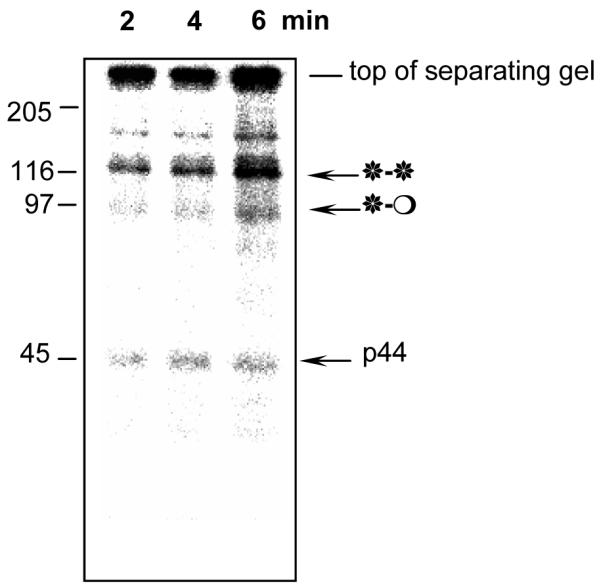
Incorporation of γ-[32P]ATP into purified ERK1 (fraction 23 from the phenylsepharose column) during in vitro phosphorylation by MEK. 0.5μl of the fraction was used per reaction as a substrate for in vitro MEK assays. Kinase reactions were carried out for 2, 4 and 6 min at 30°C, and then terminated by addition of sample buffer. Molecular mass markers are also shown. Note that radioactivity is incorporated predominantly into the band above 116kDa. The radioactivity at the top of the gel represents insoluble material that has not entered the gel.