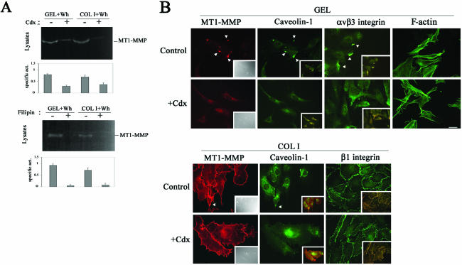
Figure 4.
Caveolae-mediated traffic is required for proper MT1-MMP activity and localization in migrating ECs. (A) Lysates from wound healing-stimulated ECs grown on GEL or COL I and pretreated or not with 1 μM cdx or 10 μg/ml filipin for 6 h were analyzed by fibrinogen zymography. The arithmetic mean and S.D. of the estimated MT1-MMP specific activity (enzymatic activity/amount of protein) from two independent experiments are shown. (B) Double staining of MT1-MMP (red) and caveolin-1 (green), or of caveolin-1 (red) and αvβ3 or β1 integrins (green) in subconfluent ECs on GEL or COL I pretreated or not with 1 μM cdx for 6 h was performed. Merged images are shown in the insets (yellow) and colocalization is marked (arrowheads). Differential interference contrast images (insets) and F-actin staining (right) of subconfluent ECs on GEL treated or not with cdx are also shown as control of cell morphology. Bar, 20 μm.

