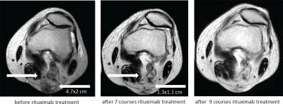
Figure 3. Complete regression of the metastases in the left popliteal region during rituximab therapy.
Popliteal metastases were screened by MRI before the start of rituximab treatment (day -28) and after 7 and 9 courses of local treatment. The popliteal bulk of metastases measured 4.7 × 2 cm before therapy and regressed to 1.3 × 1.1 cm and disappeared after 9 courses of treatment (right arrow). Left arrow indicates a solitary metastasis which completely disappeared after 7 courses. Complete remission was stable.

