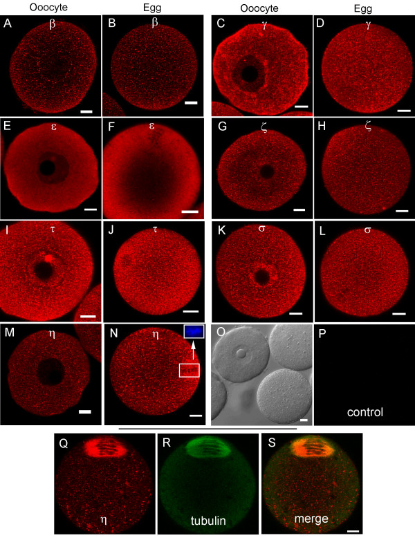
Figure 3.
Representative immunofluorescence images of 14-3-3 isoforms in oocytes and eggs isolated from adult mice. (A, B) 14-3-3β. (C, D) 14-3-3γ. (E, F) 14-3-3ε. (G, H) 14-3-3ζ. (I, J) 14-3-3 τ. (K, L) 14-3-3σ. (M, N) 14-3-3η. Confocal sections with regions of red fluorescence indicating the corresponding isoforms studied (see Methods). The inset in N shows the same egg labeled blue with Hoechst DNA stain (non-confocal image) and confirms that the darker areas in this region of the larger image are condensed metaphase II chromosomes. Control cells were included for each isoform experiment and were imaged using the same confocal settings. Representative control oocytes and eggs are shown in bright-field (O) and fluorescence (P). 14-3-3η accumulates, in part, in the meiotic spindle in eggs as shown by simultaneous labeling with 14-3-3η (Q) and tubulin (R) antibodies. These sequential scans are merged (14-3-3η + tubulin) in (S). The scale bars represent 10 μm for all images.

