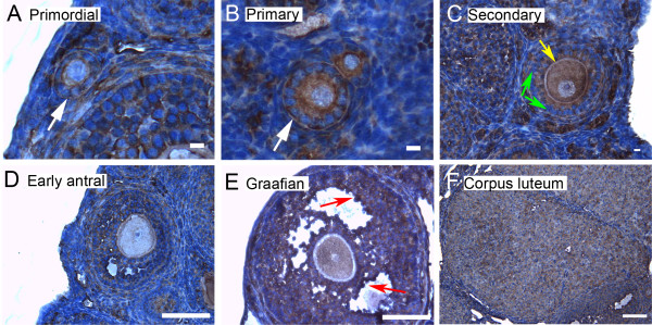
Figure 6.
Representative immunohistochemistry images of 14-3-3 ε in the different stages of follicular development in ovarian sections. (A) Primordial follicle. (B) Primary follicle. (C) Secondary follicle. (D) Early antral follicle. (E) Graafian (advanced antral) follicle. (F) Corpus luteum. White arrows indicate the primordial or primary follicles in (A and B). Note the weaker staining in mural granulosa cells in secondary follicles (C, green arrows), the more intense stain along the zona pellucida of the oocyte (C, yellow arrow), and the more intense staining in cells lining the antral cavity (E, red arrows). The scale bars represent 10 μm (A-C) or 100 μm (D-F).

