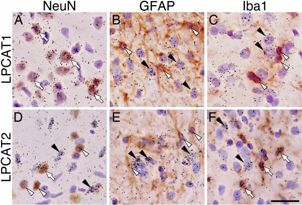
Figure 2.
LPCAT2 mRNA is increased by spinal microglia after SNI surgery. Bright-field images of combined ISHH for LPCAT1 (A-C) and LPCAT2 (D-F) with IHC for NeuN (A, C), GFAP (B, E) and Iba1 (C, F) at 3 days after SNI surgery. Open arrows indicate double-labeled cells. Arrowheads indicate single-labeled cells by ISHH (aggregation of grains), and open arrowheads indicate single immunostained cells (brown staining). Calibration bar: 20 μm.

