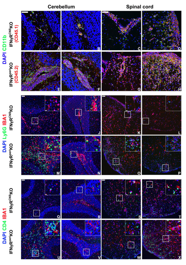
Figure 4.
Composition of inflammatory foci in the CNS of mice with EAE does not correlate with onset of atypical disease. Mice were sacrificed on day 21 pi and CNS infiltration was examined by immunohistochemistry. In IFNγRCNSKO mice CD11b+CD45.1+ infiltrating cells were usually found perivascularly or close to the meninges (A-D). In IFNγRperiKO mice, CD11b+CD45.2+ infiltrating cells were found both in perivasular spaces and deep into the parenchyma (E-H). There were few Ly6G+ neutrophils in the cerebella and spinal cords of IFNγRCNSKO mice (I-L) while the same areas of IFNγRperiKO mice were characterized by prominent neutrophilic infiltration (M-P). IFNγRCNSKO exhibited decreased CD4+ T cell parenchymal infiltration (Q-T) compared to IFNγRperiKO (U-X). Space bar = 50 μm. Inserts show magnified fields demonstrating close interactions between IBA1+ and Ly6G + (I-P) or CD4+ (Q-X) cells (space bar = 10 μm). Nuclei were visualized by DAPI staining. Two (out of 4-6) representative mice per set are shown.

