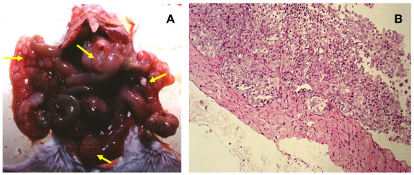
Figure 1.
Representative appearance of intraperitoneal (i.p.) tumor implants in SCID mice. A. Macroscopic view of a peritoneal metastatic tumor after i.p. injection of MFOC3. Small round metastatic foci of extensive local growth (arrowhead) are present in the peritoneum, omentum and mesenterium. B. Microscopic view of a peritoneal metastatic tumor after i.p. injection of MFOC3. MFOC3 cells (arrowhead) grew on the peritoneum.

