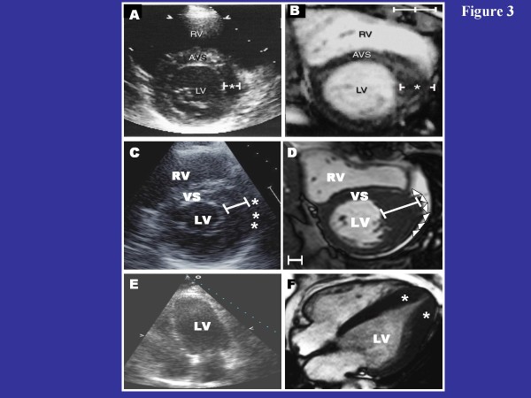
Figure 3.
CMR can identify segmental LV hypertrophy that may not be reliably visualized by two-dimensional echocardiography. (A) normal 2-D echocardiogram in a patient with a family history of HCM; (B) this same patient then underwent CMR, which reveals an area of segmental hypertrophy in the anterolateral LV wall (asterisk) consistent with a diagnosis of HCM. Reproduced with permission of American Heart Association; from Rickers C et al.;[35] (C) two-dimensional echocardiographic end-diastolic basal short-axis view demonstrates a maximal LV wall thickness of 18 mm in the anterolateral free wall consistent with the diagnosis of HCM; (D) in the same patient, an end-diastolic short-axis CMR at the same level of LV shows a focal area of massive LV hypertrophy (35 mm) in the same region of the LV wall reported to be 18 mm by echocardiography. The finding of massive hypertrophy by CMR, characterized this patient as high risk, and prompted recommendation for ICD therapy for primary prevention of sudden death. Reproduced with permission, from Maron MS et al.;[42](E) echocardiography was considered non-diagnostic; (F) in the same patient, CMR clearly demonstrates segmental hypertrophy confined to the LV apex, consistent with a diagnosis of apical HCM. Reproduced with permission, from Moon et al.[43] LV = left ventricle; RV = right ventricle; VS = ventricular septum.

