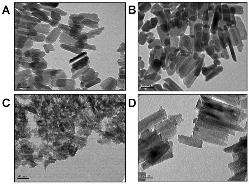
Figure 1.
Enamel crystallites in the WT mice (A and B) were well-developed and often showed a parallel organization with respect to adjacent crystallites. In contrast the AKO enamel crystallites (C) were smaller and less organized than the WT crystallites. The KOM180-87 mouse (D) showed a return to normal crystallite development in both size and with an often parallel orientation similar to that seen in the WT enamel. (TEMs all exposed at 270K magnification) (scale bar = 50 nm).

