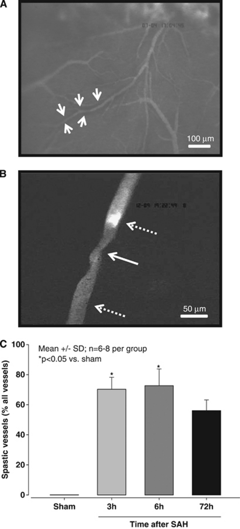
Figure 2.
(A) Microscopic overview (magnification × 100) of the middle cerebral artery (MCA) territory 3 hours after subarachnoid hemorrhage (SAH). A black halo surrounding some arterioles indicates the presence of subarachnoid blood (white arrows). (B) Pearl string-like microarteriolar spasm (solid arrow) in the cerebral microcirculation 3 hours after SAH (magnification × 250). A microthrombus occludes the proximal side of the spasm (upper dotted arrow). (C) The proportion of spastic vessels expressed as percent of all vessel segments of the ipsilateral MCA at different time points after SAH (n=6 to 8 per each time point).

