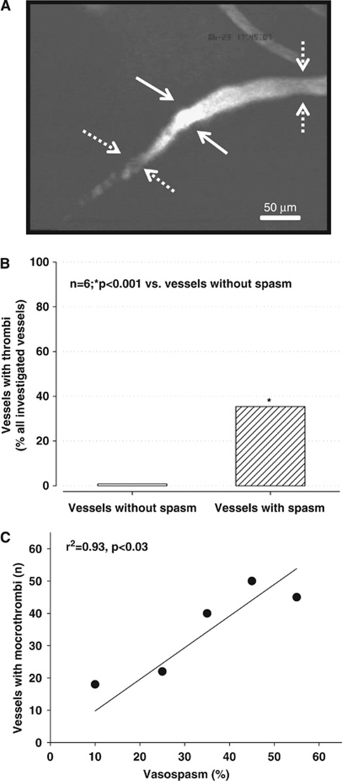
Figure 5.
(A) Example of a microvessel carrying a large thrombus (solid arrows) between two microvasospasms (dotted arrows). (B) Percentage of nonspastic (open bar) and spastic (striped bar) vessels carrying microthrombi 3 hours after subarachnoid hemorrhage (SAH). (C) Correlation between the number of microthrombi and microvasospasm severity. The more pronounced microvasospasms were the more microthrombi could be found in the corresponding vessel segments.

