Figure 2.
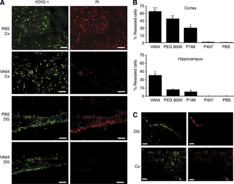
Evidence of plasma membrane resealing by Kollidon VA64 after controlled cortical impact (CCI). (A) Representative micrographs depicting cells labeled with YOYO-1 (green) and PI (red) in the cortex and DG in PBS-treated or VA64-treated mice (bottom panel). Scale bars=20 μm. (B) Quantitation of YOYO-1+/PI− (resealed) cells in the cortex and hippocampus of PBS- or polymer-treated mice (n=3 to 7 per group). Upper panel: ***P<0.05 versus all other groups; **P<0.05 versus P188, P407, and PBS; *P<0.05 versus P407 and PBS. Lower panel: *P<0.05 versus PBS and P407. (C) Representative photomicrographs showing plasmalemma resealing using intracerebroventricular administration of fluorophores and polymers (n=4 per group). It must be noted that robust membrane resealing is observed using VA64, suggesting a direct effect of VA64 on the plasmalemma of parenchymal cells. Scale bars, 100 μm. DG, dentate gyrus; PBS, phosphate-buffered saline; PI, propidium iodide. The colour reproduction of this figure is available at the Journal of Cerebral Blood Flow & Metabolism journal online.

