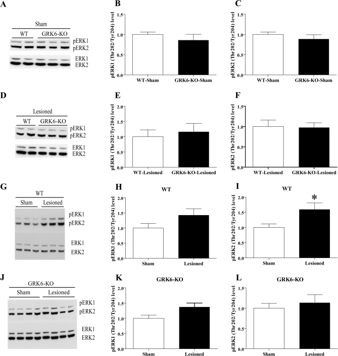
Figure 7. Levels of pERK1 and pERK2 in the striatum of unilaterally lesioned L-DOPA treated WT and GRK6-KO mice.
Western blots and densitometric analysis of relative levels of pERK1 and pERK2 were examined in extracts prepared from the striatum of WT and GRK6-KO chronically treated with L-DOPA for 21 days (n = 7–10 per group). Comparison of pERK1 and pERK2 levels in sham-operated WT and GRK6-KO mice (7A–C) and lesioned WT and GRK6-KO mice (7D–F). Comparison of pERK1 and pERK2 levels in sham-operated and lesioned WT mice (7G–I) and sham-operated and lesioned GRK6-KO mice (7J–L). Total protein levels in extracts were used as loading controls for measurement of phospho-protein levels. Data are means ± SEM; *p< 0.05, two-tailed Mann Whitney test.

