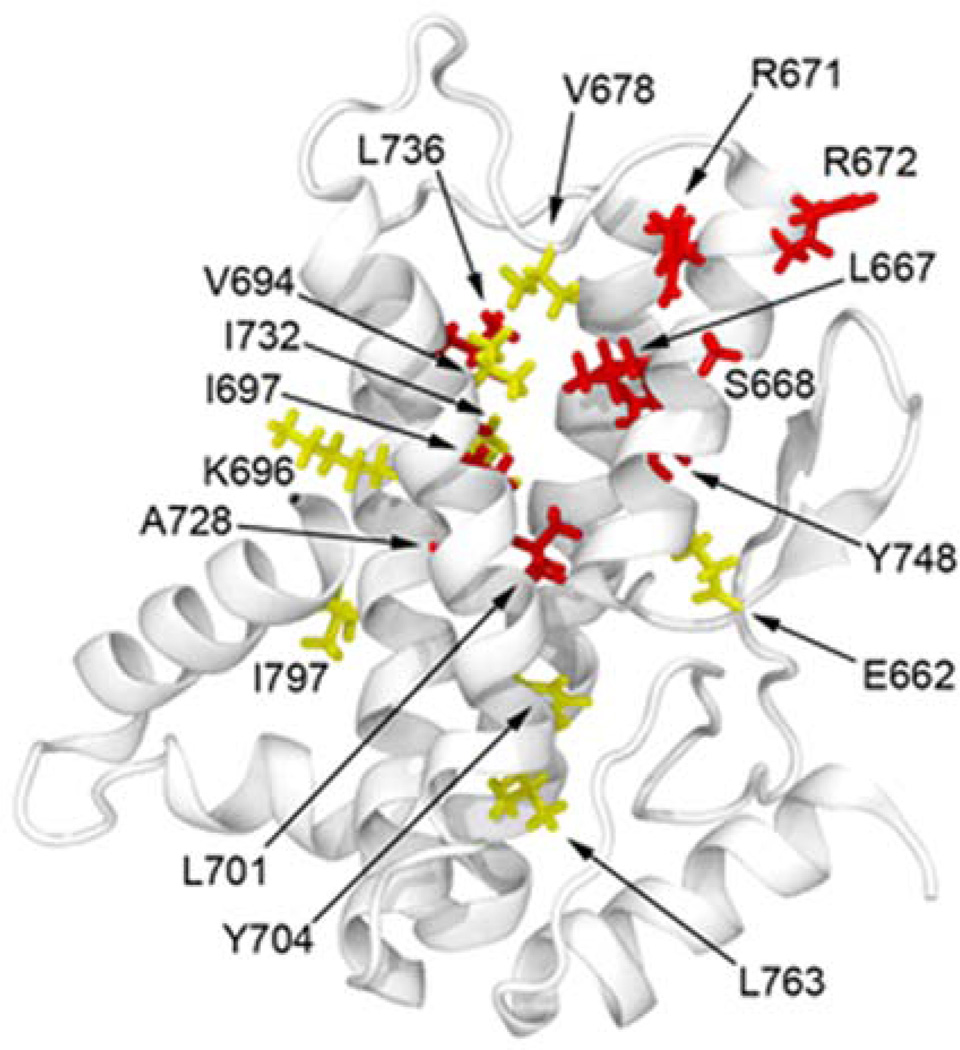
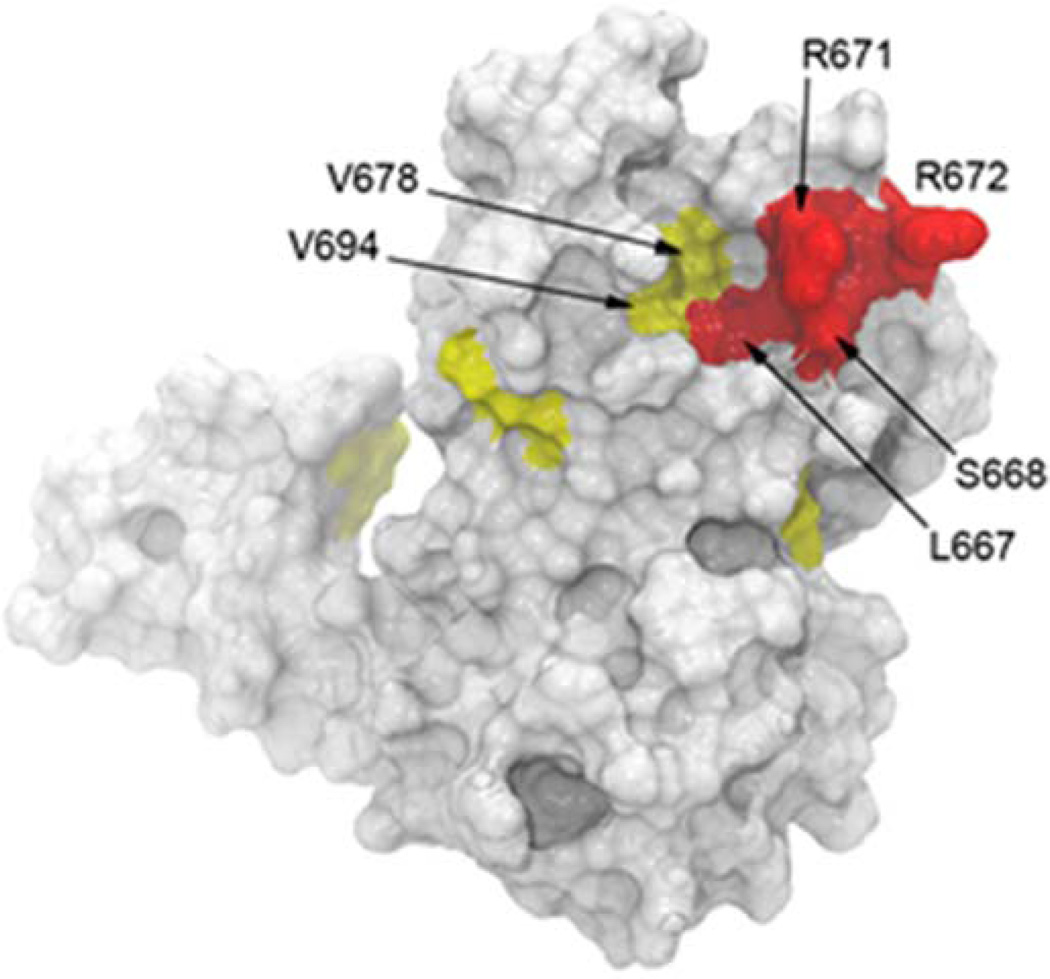
Figure 2.
Mapping of EF-neutralizing mAb EF13D on the surface of DIII. (a) Ribbon cartoon of the X-ray crystal structure of DIII (PDB accession code 1K8T) showing all the mutated amino acid residues that led to partial loss (yellow) or complete loss (red) of binding by EF13D. (b) A molecular surface representation, based on the X-ray crystal structure of DIII (PDB accession code 1K8T). The indicated amino acid residues associated with abolishing and reducing binding by mAb EF13D are shown in red and yellow, respectively. These residues are predicted to form the epitope for mAb EF13D.


