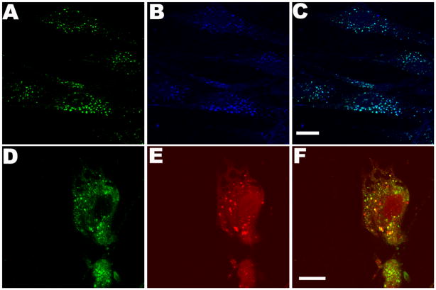
Figure 4.
Uptake and lysosomal targeting of fluorescent-labeled rhIDU into human Hurler patient fibroblast cultures. AF680 labeled enzyme (160 units) was directly applied to Hurler fibroblasts grown on coverslips in serum-free MEM and incubated for 4 hours at 37 °C and 5% CO2. (A–C) Detection of AF680 label (blue) anti-rhIDU antibodies (green) and co-localization (aqua) in Hurler cells. (D–F) Lysosomal targeting of rhIDU-AF680. Detection with anti-iduronidase antibody (green), LysoTracker Red DND-99 (red), and merge. All colors are generated by Leica LCS imaging software. Scale bars are 15 μm.

