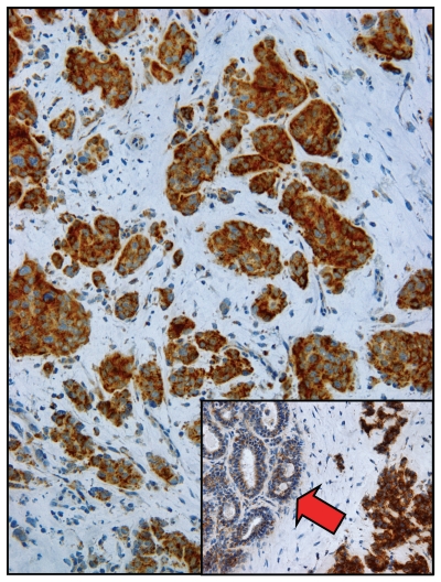
Figure 6.
Visualizing two-compartment tumor metabolism with mitochondrial activity staining. Frozen sections from human breast cancers were subjected to COX staining (brown color), which functionally detects mitochondrial activity. Note that epithelial cancer cell nests are COX-positive and, thus, are oxidative. In contrast, the tumor stroma is COX-negative and, hence, glycolytic. Inset, “normal” adjacent epithelial cells (red arrow) show substantially less COX activity as compared with epithelial cancer cells. Reproduced and modified with permission from reference 72.

