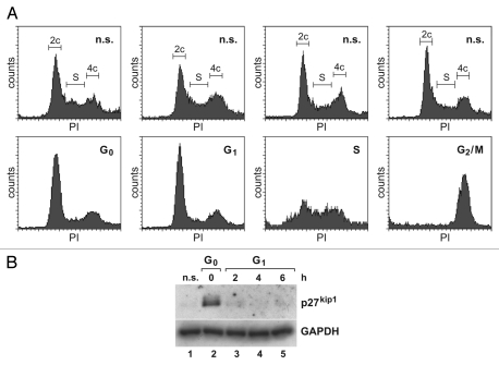
Figure 2.
Analyses of synchronized Huh7 cells. (A) Upper part: unsynchronized control cells, stained with propidium iodide (PI) and analyzed by FACS. Lower part: Huh7 hepatoma cells synchronized in the G0, G1, S and G2/M phase of the cell cycle. (B) p27kip1 western blot of G0 cells and G1 cells harvested at the times after release from contact inhibition as indicated (upper part). GAPDH was analyzed as loading control (lower part).

