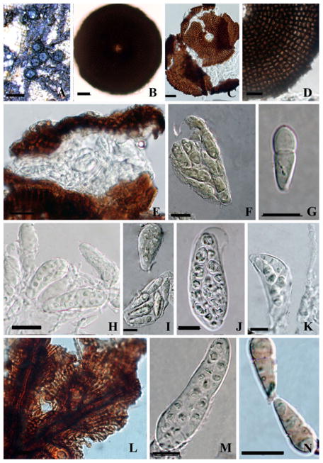
Fig. 25. Trichothyrium peristomalis (holotype of Actinopeltis peristomalis).
a Habit, appearance of ascomata on the host surface. b, c, d. Squash mount of ascoma. Note the parallel radiating arrangement of cells. E. Section of an ascoma. f, i, j, m Ascus. g, n Ascospores. Note the slightly constricted septum. H, K. Hamathecium. Note the pseudoparaphyses L. Superficial hyphae Note forming thalli. Scale bars: A=200 μm, B, C, I=20 μm, D–N=10 μm

