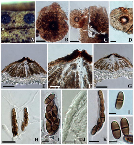
Fig. 4. Arnaudiella caronae (PDD 54414).
a Habit, appearance of ascomata on the host surface. b, c, d Squash mount of ascoma. Note the scutate structure and radiating arrangement of hypha. e–g Section of an ascoma. Note the peridium and F note ostiole. h–k Ascus Note with a pedicel and pseudoparaphyses in Melzer’s reagent. L, M. Ascospores. Scale bars: A=500 μm, B, C, E, G=50 μm, D, J, K= 10 μm, F, H=20 μm, I, L, M=5 μm

