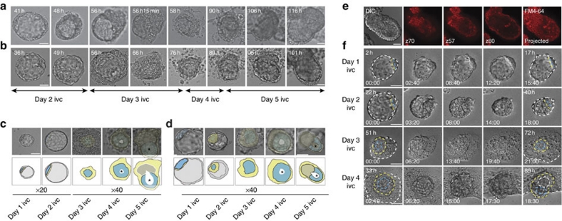
Figure 1. In vitro culture of blastocysts on collagen-coated polyacrylamide matrices allows successful development to the egg cylinder stage.
(a, b) DIC time-lapse observations of in vitro development of two representative blastocysts on collagen-coated polyacrylamide gels covalently bound to a glass-bottomed dish. Each panel shows a single embryo continuously observed from the second day of in vitro culture (ivc), until the fifth day. Panels (a) and (b) show selected frames from Supplementary Movies 1 and 2, respectively. Elapsed developmental time from the point at which the blastocyst was placed on the gel is shown at the top of each frame. Scale bars represent 50 μm. (c, d) Development of two further representative embryos in vitro and observed using a ×40 objective. DIC images are shown alongside schematics showing different tissues: epiblast, blue; primitive/visceral endoderm, yellow; trophectoderm/ExE, grey; the pro-amniotic cavity is highlighted by an asterisk. The overlying trophectoderm is omitted from the schematics of the third and fourth day of in vitro culture, for clarity. Development of the embryo shown in (c) is presented in Supplementary Movie 3. (e) On day 5 of in vitro culture, the embryo shown in (d) was removed from the gel and stained with the lipophilic dye FM4-64. DIC, selected confocal stacks and projected images are shown. (f) DIC time-lapse observations during 4 days of development in vitro. A representative embryo is shown for each day of culture. Elapsed developmental time from the point at which the blastocyst was placed on the gel is shown at the top of the first and the last frame presented for each day. Time points at the bottom of each still represent individual time frames from time-lapse observations on the indicated day. Embryonic and extra-embryonic structures are indicated as follows: primitive endoderm (blastocyst) and visceral endoderm (egg cylinder), yellow-dashed line; trophectoderm (blastocyst) and ExE (egg cylinder), white-dashed line; epiblast, blue-dashed line. Scale bars represent 50 μm.

