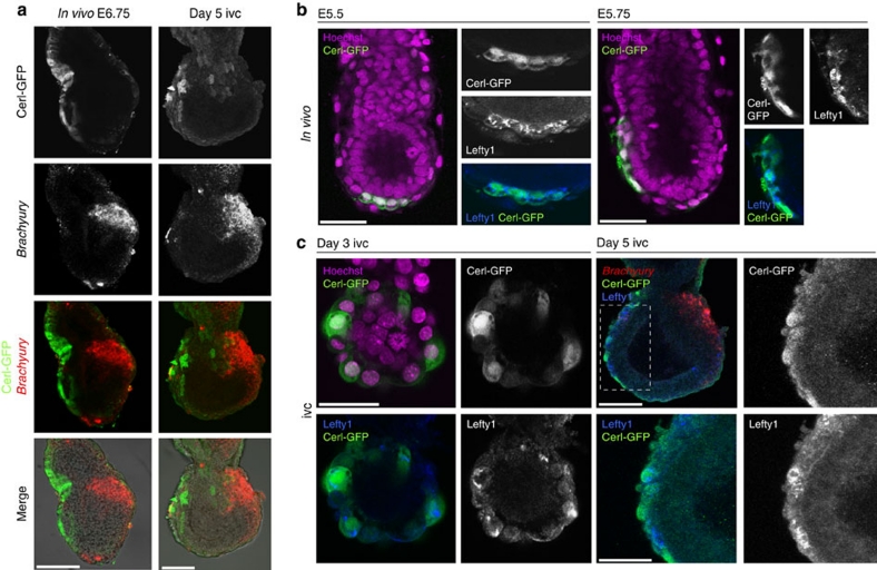
Figure 3. Spatial expression of molecular markers of development of in vitro cultured embryos.
(a) Fluorescent in situ hybridisation for Brachyury with co-immunostaining for GFP in E6.5 in vivo- (upper panels) and day 5 in vitro-cultured (lower panels) Cerl-GFP transgenic embryos. AP patterning is faithfully recapitulated in five out of seven embryos. Scale bars represent 100 μm. (b) Co-immunostaining for Lefty1 and GFP in E5.5 and E5.75 Cerl-GFP transgenic embryos. Expression of the reporter gene is observed in the same region as another AVE-specific protein, Lefty1. Scale bars represent 50 μm. (c) Co-immunostaining for Lefty1 and GFP in day 3 and day 5 in vitro-cultured Cerl-GFP transgenic embryos. The expression of Lefty1 is restricted to the GFP-positive domain throughout development. Scale bar represents 50 μm. On the fifth day of in vitro culture (right-hand side panels), Brachyury expression is detected by FISH opposite to Lefty1, and Cerl-GFP domain is detected by co-immunofluorescence. Scale bars represent 100 μm.

