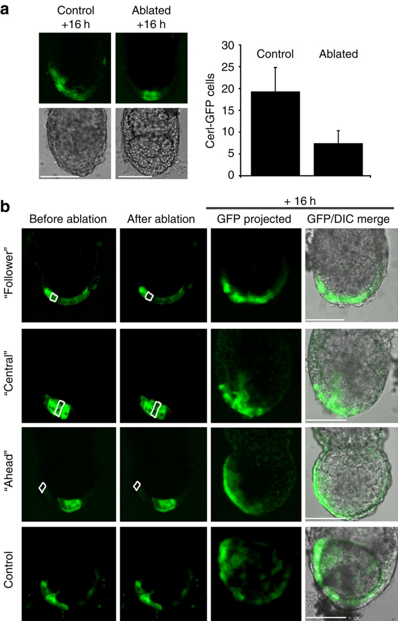
Figure 7. AVE migration requires leading AVE cells.
(a) Projected confocal images of control embryos (left panels) and an embryo in which the leading AVE cell was ablated by laser-irradiation (right panels, Supplementary Movie 8) 16 h after ablation. Migration of the Cerl-GFP expressing cells is prevented when the leading AVE cell is ablated. Numbers of Cerl-GFP expressing cells in control and leading cell-ablated embryos were quantified 16 h after ablation: Ablation of leading cells prevents expansion of the Cerl-GFP-expressing domain. (b) Consequences of cell ablation for AVE migration and expansion: cell ablation immediately behind the leading cell, in the central section of the AVE, and in a region of visceral endoderm anterior to the AVE. Images of Cerl-GFP expression are taken immediately before and after ablation, and 16 h later. Scale bars represent 100 μm.

