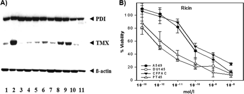
FIGURE 5.
PDI and TMX expression and reduction of ricin in human cell lines. A, immunoblot identification of TMX and PDI in human cell lines extracts. Lane 1, A549 (respiratory epithelia); lane 2, DU145 (prostate cancer); lane 3, LNCaP (prostate cancer); lane 4, T3M4 (pancreatic cancer); lane 5, GER (pancreatic cancer); lane 6, PACA44 (pancreatic cancer); lane 7, MiaPaCa (pancreatic cancer); lane 8, CFPAC-1 (pancreatic cancer); lane 9, PT45 (pancreatic cancer); lane 10, Jurkat (T cell leukemia); lane 11, THP-1 (human monocyte). Human β-actin was used as a control to determine the relative amount of protein for each cell line. B, A549, DU145, PT45, and CFPAC-1 cells were incubated with ricin at various concentrations. The vitality was tested after 48 h by XTT assay as described above.

