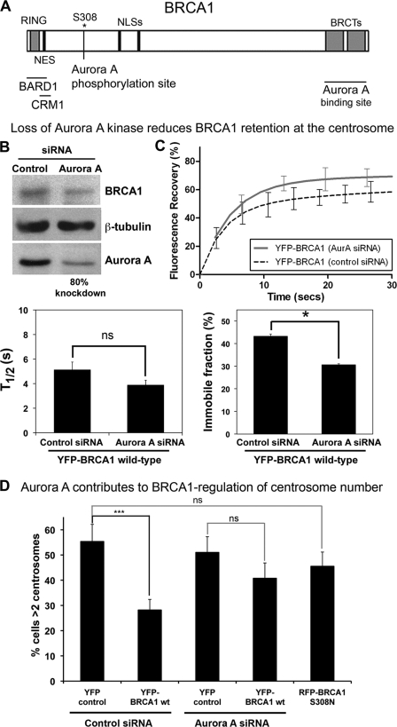
FIGURE 8.
Aurora A kinase regulates BRCA1 dynamics and function at the centrosome. A, diagram showing sites where Aurora A kinase binds (BRCT domain) and phosphorylates (Ser-308) BRCA1. B, Western blot analysis to confirm the Aurora A knockdown using siRNA transfection in MCF-7 cells. C, FRAP analysis was performed on live MCF-7 cells transfected with YFP-BRCA1 (and RFP-pericentrin C241 as marker) after transfection with either control or Aurora A siRNA. Corresponding recovery curves are shown, indicating the immobile and mobile fractions and percentage increase in mobility after Aurora A silencing. The fast-phase half-time (t½ ± S.E. (error bars)) and immobile fraction (percentage ± S.E.) are shown for each construct, analyzing 10–15 cells/sample. D, functional assay. pYFP-BRCA1 WT was transfected into HCC1937 breast cancer cells after treatment with Aurora A or control siRNA. pRFP-BRCA1 (S308N) was also transiently expressed in HCC1937 cells. The cells were then irradiated (10 Gy of IR) and left to recover for 48 h. After fixation with acetone/methanol, cells were immunostained with anti-γ-tubulin antibody and scored for cells displaying centrosome amplification. Scoring results were obtained from at least three independent experiments with at least 100 cells scored (mean ± S.D. (error bars)). ***, p < 0.001. ns, not significant.

