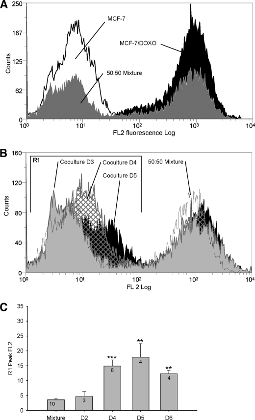
FIGURE 2.
Analysis by flow cytometry of P-gp transfers in co-cultures. Membrane P-gp content was assessed by flow cytometry in the FL2 channel after direct immunolabeling with PE-UIC2. A, cell membrane P-gp content was quantified in parental MCF-7 (open black histogram), in resistant MCF-7/Doxo (solid black histogram), or in an extemporaneous mixture of 50:50 MCF-7:MCF-7/Doxo (solid gray histogram). B, membrane P-gp content was quantified in co-cultures. A 50:50 MCF-7:MCF-7/Doxo cell mixture was seeded on culture dishes at day 0 and trypsinized for increasing co-culture durations. P-gp distribution in an extemporaneous mixture of 50:50 MCF-7:MCF-7/Doxo was superimposed for comparison (open black histogram). Results obtained at days 3 (solid gray histogram), 4 (hatched histogram), and 5 (solid black histogram) show a progressive shift to the right of the peak initially corresponding to sensitive MCF-7 low P-gp expression. C, shown is a statistical comparison of the peak FL2 fluorescence in region R1 after different durations of co-culture (day D1 to D6), expressed as the mean ± S.E. of the replicate numbers indicated in the bars. Results significantly different from the mixture are indicated (*, p < 0.05; **, p < 0.01; ***, p < 0.001). The maximum P-gp transfer was observed at day 5.

