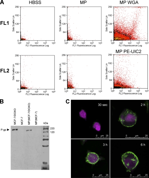
FIGURE 5.
Characterization of microparticles released by MCF-7/Doxo. MPs were extracted and purified from MCF-7/Doxo cultures as described under “Experimental Procedures.” A, MPs were detected in flow cytometry by their capability to diffract the laser light source and to give an increased side scatter signal (middle dot plots) by comparison to a sample of pure HBSS (left dot plots). Glycoconjugates in MP membranes were revealed with Alexa Fluor 594 conjugated-WGA and analyzed in the FL1 channel (top right dot plots). P-glycoprotein was detected in MPs by PE- UIC2 labeling in the FL2 channel (bottom right dot plots). B, total P-gp content of MPs was studied by Western blot using c219 as anti-P-gp primary mAb. P-gp was clearly detected in protein extracts from MCF-7/DOXO (lane 1) and from MPs extracted from MCF-7/DOXO cell cultures (lane 3) but not in extracts from MCF-7 (lane 2) and from MPs extracted from MCF-7 cell cultures. C, detection MP binding to MCF-7 cells is shown. CellTracker Violet-tagged MCF-7 cells were exposed to WGA-labeled MPs extracted from MCF-7/DOXO and imaged by confocal microscopy. Intensity of WGA labeling increased at the membrane of MCF-7 with time of incubation with MPs from 30 s to 6 h.

