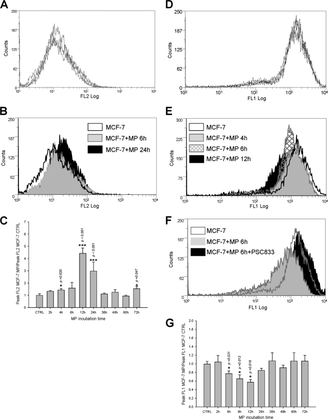
FIGURE 6.
Transfers of P-gp and efflux activity to sensitive MCF-7 exposed to MPs extracted from MCF-7/DOXO. A, repeatability of P-gp distributions in MCF-7, measured by flow cytometry after direct immunolabeling by PE-UIC2, is shown. Histograms from five independent cultures were superimposed. B, membrane P-gp content in MCF-7 (open black histogram) treated for 6 h (solid gray histogram) or for 24 h (solid black histogram) with MPs extracted from MCF-7/DOXO increased with time of exposure. C, relative increase of membrane P-gp content in MCF-7 cells treated with MPs extracted from MCF-7/DOXO was transient, significant after 4 h, and peaked after 12 h of exposure. Results are expressed as the mean ± S.E. of n = 2–10 replicates. D, repeatability of P-gp activity distributions in MCF-7, assessed by their ability to efflux intracellular fluorescence after loading with calceinAM, is shown. Histograms from five independent cultures are superimposed. E, P-gp activity in MCF-7 cells (open black histogram) treated for 4 h (solid gray histogram), 6 h (hatched histogram), or for 12 h (solid black histogram) with MPs extracted from MCF-7/DOXO increased with time. F, P-gp activity transferred to MCF-7 by a 6-h exposure to MPs extracted from MCF-7/DOXO was blocked by the specific P-gp antagonist PSC833 (20 μm). G, relative increase of P-gp activity, expressed as a drop of intracellular fluorescence, in MCF-7 treated with MPs extracted from MCF-7/DOXO was transient, significant after 4 h, and peaked after 12 h of exposure. Results are expressed as the mean ± S.E. of n = 2–10 replicates. Results significantly different from control are indicated (*, p < 0.05; **, p < 0.01; ***, p < 0.001).

