FIGURE 7.
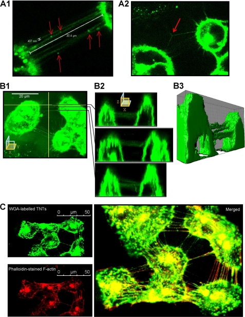
Characterization of TnTs connecting MCF-7 cells in culture. A1, Alexa Fluor 594 conjugated-WGA-stained MCF-7 cells were analyzed by live cell confocal microscopy. Cells are connected to surrounding cells by numerous ultrafine membrane extensions, namely TnTs, having a diameter inferior to 0.5 μm and a length of up to one cell diameter. WGA-stained organelles are seen at several points in TnTs (arrows). A2, branched TnTs were rarely observed. B1–B2, some TnTs were selected for x-z sections. TnTs did not contact the substrate. B3, shown is a three-dimensional reconstruction. C, fixed MCF-7 cells were stained with WGA (top left image) and TRITC-phalloidin (bottom left image). The merged image displays colocalization of WGA and TRITC-phalloidin staining, indicating that F-actin is a major component of TnTs (right image).

