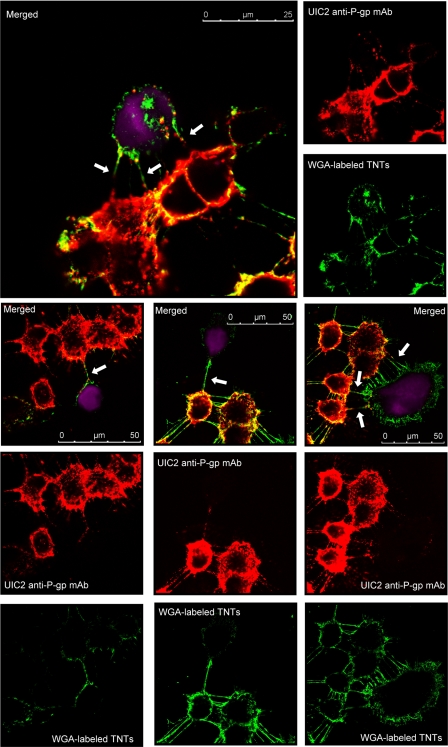
FIGURE 8.
Immunodetection of P-gp in TnTs connecting MCF-7/Doxo to MCF-7. Sensitive MCF-7 were tagged with CellTracker Violet (ctvMCF-7, violet fluorescence), before co-culture. Mixtures of 50:50 ctvMCF-7:MCF-7/Doxo were co-cultured during 4 days on glass coverslips. P-gp was detected in fixed cells by using a non-conjugated UIC2 mAb as primary anti-P-gp mAb, revealed with a Texas Red-conjugated anti-mouse IgG2a γ2a (red fluorescence), and TnTs were labeled with WGA (green fluorescence). The figure presents different images of P-gp and TnTs localization within the co-cultures. Merged images present P-gp-containing TnTs connecting resistant MCF-7/Doxo to parental ctvMCF-7 cells (arrows).

