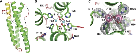
FIGURE 2.
Crystal structure of FBXL5 hemerythrin domain. A, ribbon representation of the hemerythrin domain from H. sapiens FBXL5 (residues 5–159; PDB code 3V5X). Helices are shown in green, and loops are in shown in red. The additional fifth C-terminal helix found is shown in yellow. The dotted lines represent the disordered residues 75–76 and 81–83. B, the first coordination sphere iron ligands of the FBXL5 Hr diiron center are shown as green sticks, and the conserved second coordination sphere iron ligands are shown as pink sticks. Iron ligands and hydrogen bonds are shown as dotted black lines. C, diiron center of FBXL5 Hr Native 1, monomer A with superimposed Fo − Fc simulated annealing omit electron density map (gray, contoured at 3.0 σ) calculated for the bridging oxygen (red) and the side chains (green) of the first coordination sphere (atoms beyond the C-β for the amino acids were omitted). An anomalous difference Fourier electron density map (red, contoured at 15.0 σ) is shown for the iron atoms (cyan).

