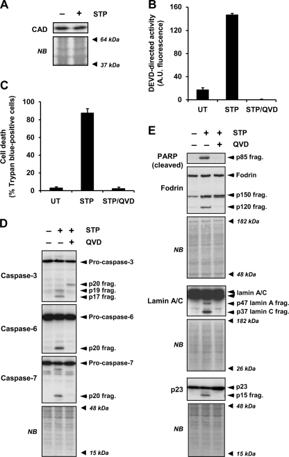
FIGURE 3.
STP-induced apoptosis in SK-N-AS cells is a caspase-dependent process. A, SK-N-AS cells were left untreated (−) or treated (+) for 24 h with 1 μm STP. Total extracts were obtained, and DFF40/CAD protein levels were analyzed by Western blot. B and C, SK-N-AS cells were left untreated (UT) or treated with 1 μm STP alone or in the presence of the pan-caspase inhibitor q-VD-OPh (20 μm) (STP/QVD). DEVD-like activity (B) or cell viability by trypan blue exclusion assay (C) was performed after 24 h of treatment. D and E, SK-N-AS cells were left untreated (−) or treated (+) with 1 μm STP alone or plus q-VD-OPh (20 μm) for 6 h. Pro-caspase-3, -6, and -7 processing in their active fragments (D) and cleavage of poly(ADP-ribose) polymerase (PARP (cleaved)), α-Fodrin, lamin A/C, and p23 co-chaperone (E) were analyzed by Western blot. The apparent molecular mass of the specific fragments is indicated on the right of the panels. The membranes were stained with naphthol blue (NB) to assess equal loading. A.U., fluorescence arbitrary units.

