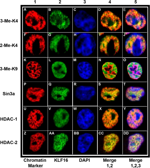
FIGURE 9.
KLF16 compartmentalization to euchromatin with chromatin cofactors in cultured endometrial cells. For coimmunolocalization of KLF16 in uterine cells, representative images are shown. All cells were stained with DAPI (blue, column 3, panels C, H, M, R, W, and BB) to visualize the DAPI-light euchromatic and DAPI-intense heterochromatic regions. All cells were also stained with anti-KLF16 (green, column 2, panels B, G, L, Q, V, and AA). For chromatin colocalization, cells were additionally stained with specific monoclonal antibodies (red, column 1, panels A, F, K, P, U, and Z) to either euchromatic markers trimethyl H3K4 or dimethyl H3K4 (panels A–E and F–J, respectively) or a heterochromatic marker trimethyl H3K9 (panels K–O). Overlay of corresponding KLF16 and individual chromatin markers (column 4, panels D, I, N, S, X, and CC) and of KLF16, individual chromatin markers and DAPI (column 5, panels E, J, O, T, Y, and DD), respectively, is shown in the figure, in columns 4 and 5 are labeled merge 1,2 and merge 1, 2, 3, respectively. KLF16 preferentially colocalized with euchromatin markers (A–J) compared with the heterochromatic marker (K–O). To evaluate colocalization with the Sin3a corepressor complex, cells were stained with monoclonal antibodies to Sin3a, HDAC1, and HDAC2 (panels P–T, U–Y, and Z–DD, respectively). KLF16 extensively colocalized with Sin3a, HDAC1, and HDAC2 (P–DD).

