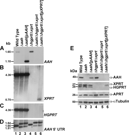
FIGURE 3.
Southern and Western blot analysis of the Δaah and Δaah/Δhgprt/Δxprt mutants. Total genomic DNA from wild type (lane 1), Δaah (lane 2), Δaah[pAAH] (lane 3), Δhgprt/Δxprt (lane 4), Δaah/Δhgprt/Δxprt (lane 5), and Δaah/Δhgprt/Δxprt[pXPRT] (lane 6) parasites was digested with EcoRI or SmaI and BamHI, fractionated on a 0.8% agarose gel, and blotted onto nylon membranes. The blot was hybridized under stringent conditions with a probe to the full-length ORFs of AAH (A), XPRT (B), HGPRT (C), or the AAH 5′ UTR (D). The 1,650-bp wild type AAH EcoRI fragment encompasses 765 bp of the AAH ORF and 885 bp of the AAH 5′ UTR (A, lanes 1 and 4) and the 4.3-kb EcoRI fragment encompasses the full-length coding sequences of both XPRT and HGPRT (B and C, lanes 1–3). The coding sequences of AAH and XPRT were excised from the pXG-PHLEO-AAH and pXG-BSD-XPRT episomes, respectively, by digestion with SmaI/BamHI and present as 1,089-bp (A, lane 3) and 726-bp (B, lane 6) fragments, respectively. E, lysates of exponentially growing wild type (lane 1), Δaah (lane 2), Δaah [pAAH] (lane 3), Δhgprt/Δxprt (lane 4), Δaah/Δhgprt/Δxprt (lane 5), and Δaah/Δhgprt/Δxprt[pXPRT] (lane 6) parasites were analyzed by immunoblotting with monospecific polyclonal antisera to AAH, XPRT, HGPRT, and APRT, as shown. The amount of protein loaded onto each lane was normalized using commercially available mouse anti-α-tubulin monoclonal antibody.

