FIGURE 4.
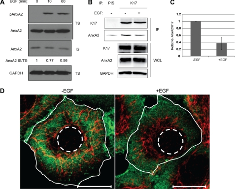
EGF induces dissociation of K17 and AnxA2. A, growth factor-deprived A431 cells were treated with 100 ng/ml EGF for the indicated time periods. Whole cell lysates were processed for Triton solubility, and immunoblotting was performed with antibodies against the indicated proteins. AnxA2 signal intensities were quantified using ImageJ. B, growth factor-deprived A431 cells were treated with (+) or without (-) 100 ng/ml EGF for 30 min. Immunoprecipitation was performed with anti-K17 antibody (K17) or preimmune serum (PIS) as a control, and immunoblotting was performed with antibodies against the indicated proteins. C, AnxA2 and K17 signal intensities from coimmunoprecipitation were quantified using ImageJ, and relative AnxA2/K17 is shown. D, growth factor-deprived A431 cells were treated with (+EGF) or without (-EGF) 100 ng/ml EGF for 30 min and then immunostained with anti-K17 (red) and anti-AnxA2 (green) antibodies. The plasma (solid lines) and nuclear (dotted lines) membranes are shown. Scale bar = 20 μm.

