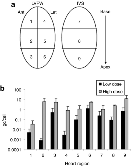
Figure 1.
Taqman analysis of vector genome distribution in the heart. (a) For analysis, biopsies were collected from the LVFW and IVS from base to apex as depicted. (b) Biodistribution of vector genomes throughout the heart in individual high- and low- dose GRMD canines. Error bars represent standard deviations. Ant, anterior; gc, genome copies; GRMD, golden retriever muscular dystrophy; IVS, interventricular septum; Lat, lateral; LVFW, left ventricular free wall.

