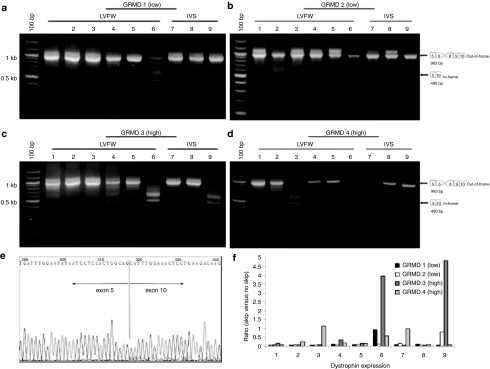
Figure 3.
RT-PCR analysis of exon skipping in GRMD canines treated with rAAV6-U7-ESE6/8. The GRMD mutation causes loss of exon 7, producing an out-of-frame transcript. Treatment with ESE6/8 will cause splicing of exon 5 to exon 9 or exon 10 (as cryptic skipping of exon 9 is a well-described phenomenon), both of which are in-frame transcripts. For analysis, biopsies were collected from the LVFW and IVS from base to apex as depicted in Figure 1a. Representative gel images are shown from GRMD canines treated with (a,b) 1.8 × 10¹² gc/kg of rAAV6-U7-ESE6/8 and euthanized 13 months later (n = 3), and (c,d) 1.4 × 10¹³ gc/kg of rAAV6-U7-ESE6/8 and euthanized 13 months later (n = 2). Samples were considered positive for exon skipping if they contained the 490 bp band indicative of in-frame splicing of exon 5 to exon 10, as depicted in the diagram adjacent to each gel image. Samples were considered negative for exon skipping if they contained only the 953 bp band representing loss of exon 7 secondary to the GRMD mutation. Note that a greater number of samples were positive for exon skipping following treatment with high-dose rAAV6 compared to lower doses of rAAV6. (e) Sequencing of the 490 bp band confirmed splicing of exon 5 to exon 10, which produces an in-frame transcript. (f) Quantitative analysis confirmed that the ratio of in-frame skipped product to unskipped product was higher in canines treated with high-versus low-dose rAAV6. AAV, adeno-associated virus; GRMD, golden retriever muscular dystrophy; IVS, interventricular septum; LVFW, left ventricular free wall; rAAV76, recombinant AAV6; RT-PCR, reverse transcription-PCR.

