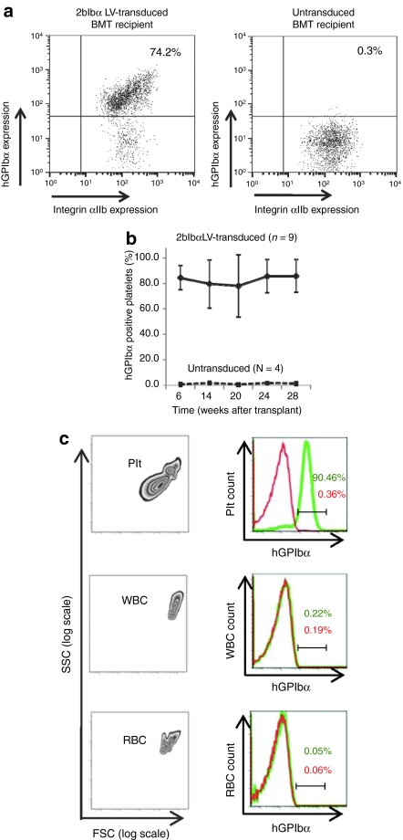
Figure 2.
Flow cytometric analysis of human GPIbα (hGPIbα) expression in bone marrow transplantation (BMT) recipients. (a) Expression of hGPIbα in the platelets of 2bIbα lentiviral vector (LV) transduced (upper panel) and untransduced (lower panel) hematopoietic stem cells (HSC) recipients were analyzed by flow cytometry. The platelet population was gated with anti-mouse CD41/integin αIIb mAb and hGPIbα expression was analyzed using AlexaFluor 647 labeled anti-hGPIbα mAb (AP1). (b) Expression of hGPIbα was monitored for 28 weeks after BMT and average expression (percentage of hGPIbα-positive platelets) in 2bIbα LV-transduced HSC recipients (n = 9) was plotted at each time point. Untransduced controls (n = 4) were analyzed in parallel each time. Data is expressed as the mean ± SD. (c) Platelet-specific expression of hGPIbα. Entities exhibiting the forward (FSC) and side (SSC) scattering properties of platelets (Plt), white blood cells (WBC), and red blood cells (RBC) from whole blood of 2bIbα LV-transduced HSC recipients (left columns) were gated to analyze hGPIbα expression on the various blood cell populations. Right columns show histograms of hGPIbα expression in transduced (green) and untransduced (red) HSC recipients. Only platelets from transduced recipient display hGPIbα on their surface.

