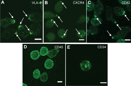
Figure 1.
A polarized membrane domain is found on normal human HSPCs. (A-E) Immunofluorescence labeling of human MPB CD34+ cells from a normal volunteer with antibodies for VLA-4 (A), CXCR4 (B), and CD82 (C) shows a polarized distribution of the membrane proteins on the majority of the cells, whereas CD45 (D) and CD34 (E) are distributed uniformly on the plasma membrane. These images are representative of the polarized molecule phenotypes observed in more than 100 CD34+ cells. Scale bars in panels A through E equal 5 μm.

