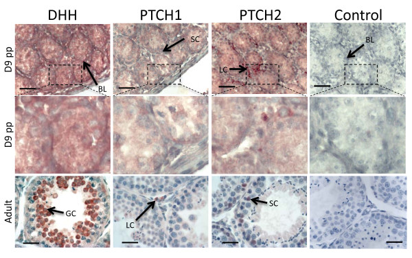
Figure 5.
Immunohistochemistry of DHH, PTCH1 and PTCH2 in the tammar wallaby testis at key developmental time points. Red/brown staining indicates protein distribution while the heamatoxalin counterstain appears blue. It is important to note that DHH is a highly secreted molecule and staining does not imply cell of origin. DHH staining was most intense at the basal lamina (BL). In the adult staining is concentrated in the develop spermatocytes (GC). At day D9pp PTCH1 was present within the Sertoli cells (SC) while PTCH2 was predominant in the Leydig cells (LC). This expression profile is reversed in the adult testis with PTCH1 found predominantly in the Leydig cells, while PTCH2 was predominate in the Sertoli cells. Scale bars indicate 40 μm, controls show immunohistochemistry with the primary antibody omitted.

