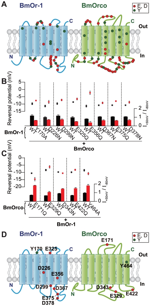
Figure 1. Mutations in BmOr-1 and BmOrco that affected reversal potential and rectification index.
(A) Schematic of the location of amino acids in BmOr-1 and BmOrco that were mutated in this study. Transmembrane domains were predicted using the PHDhtm algorithm [48]. (B,C) Reversal potential (top) and rectification index (bottom) of oocytes expressing mutant BmOr-1 with wild type (WT) BmOrco (B) or WT BmOr-1 with mutant BmOrco (C). Black and red bars and symbols represent WT and mutants, respectively. Only mutants with a significant effect on both reversal potential and rectification index are depicted (unpaired Student's t-test, mutant vs. WT p<0.05). Data on remaining mutants can be found in Figure S2B. Data are shown as mean ± S.E.M., n = 8–10. Bombykol was applied at the concentration of 1 µM to each oocyte. (D) Schematic showing the eight mutations in BmOr-1 and five mutations in BmOrco that affected both rectification index and reversal potential (see also Figure S2B,S3A).

