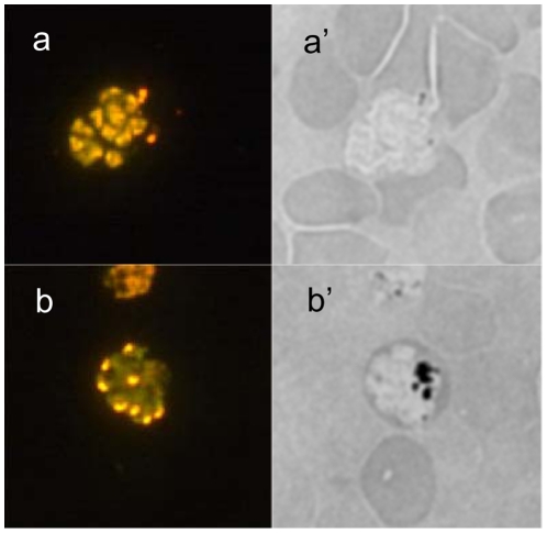
Figure 4. Epifluorescence microscopy.
Y-ART3 line schizont stained with acridine orange (a) showed a typical multiple nuclei stage but no malaria pigment inclusion could be observed in a bright field (a'). Y-control line schizont stained with acridine orange (b) showed also a typical multiple nuclei stage but there is a large amount of malaria pigment also called hemozoin (the black inclusion) observed in a bright field (b').

