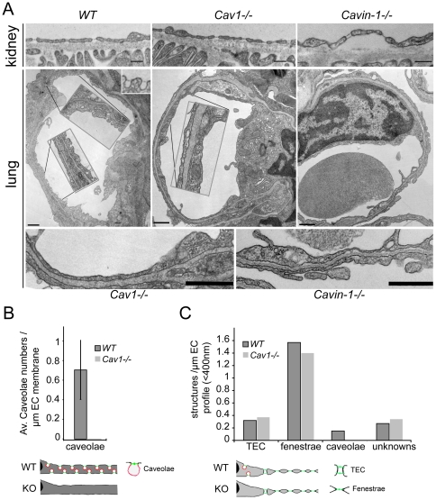
Figure 2. Protein level of PV1 correlates with the number of structures capable of forming diaphragms in vivo.
A) Electron micrographs of capillary ECs of the kidneys (top panels) and lungs (middle and bottom panels) of WT, Cav1−/− and cavin-1−/− mice, as indicated. TEC and fenestrae are present in the kidneys of WT, Cav1−/− and cavin-1−/− mice (top panels). (Middle and bottom panels) Caveolae with stomatal diaphragms are present in the lungs of WT and absent in Cav1−/− and cavin-1−/− mice (middle panel). Insets in middle panels are a 2-fold magnification of the noted stretches of ECs. Bottom panels are a 3-fold magnification of ECs of Cav1−/− (left) and cavin-1−/− (right). Bars −200 nm. B) Morphometric analysis of the number of lung endothelial caveolae in WT and Cav1−/− mice demonstrating the absence of caveolae in the latter. C) Morphometric analysis of the numbers of kidney endothelial TEC, fenestrae and caveolae in WT and Cav1−/− mice.

