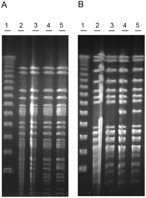
Figure 2. PFGE of outbreak associated isolates.
PFGE of E. coli O103:H25 NOS (lane 2), NVH-847 (lane 3), NVH-848 (lane 4), and NVH-760 (lane 5). Lambda ladder is used as marker (lane 1). Digestion with XbaI (A) showed indistinguishable PFGE patterns, while digestion with AvrII (B) exposed a difference between the stx2-positive and the stx-negative isolates.

